
HIGH RESOLUTION SONOGRAPHY IN ROTATOR CUFF TEAR
Following set of representative shoulder ultrasound images have been submitted by Dr Subhash Tailor, MD-Consultant Radiologist at Bhilwara.
CASE 1 - PARTIAL SUPRASPINATUS TENDON ARTICULAR FIBRES TEAR
A 45 yrs male with h/o trauma right shoulder , painful arc syndrome & inability to abduct right shoulder .
AT US – focal inhomogenicity with hyopoechoic defect in articular fibres of right supraspinatus tendon , & adjacent GT cortical iiregularity , s/o partial supraspinatus tear. [ see figure 1 ]

Figure 1- Focal inhomogenicity supraspinatus with adjacent greater tuberosity cortical irregularity s/o -Partial tear articular fibres
CASE 2 - FOCAL FULL THICKNESS TEAR SUPRASPINATUS TENDON
A 60 years female with h/o trauma with pain & inability to abduct shoulder
AT US - focal fluid filled defect in supraspinatus tendon involving full thickness with mild subdeltoid bursal effusion . [ see figure 2]

Figure 2 – shows echolucent full thickness defect in supraspinatus tendon with mild subdeltoid bursal effusion
CASE 3 - FULL THICKNESS SUPRASPINATUS TENDON TEAR WITH COMPLETE RETRACTION , BARE TUBEROSITY SIGN
A 75 years old man with h/o fall , & was unable to abduct shoulder
AT US - There is complete absence or nonvisualsation of supraspinatus tendon at its position , s/o Naked/ Bare tuberosity sign . In this case deltoid muscle is directly resting on humeral head . [see Figure 3]
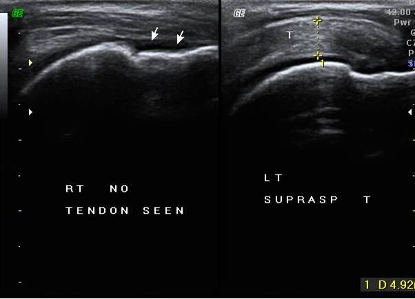
Figure 3 – Nonvisualisation of supraspinatus tendon ,& deltoid muscle is directly resting on humeral head – Bare /Naked tuberosity sign
HIGH RESOLUTION SONOGRAPHY IN ROTATOR CUFF TEAR
 Reviewed by Sumer Sethi
on
Sunday, April 15, 2012
Rating:
Reviewed by Sumer Sethi
on
Sunday, April 15, 2012
Rating:
Reviewed by Sumer Sethi
on
Sunday, April 15, 2012
Rating:
Sumer Sethi

Unique blend of academic excellence and entrepreneurship, heading leading firms in India- Teleradiology Providers, pioneering company providing teleradiology services and DAMS (Delhi Academy of Medical Sciences) Premier test preparation institute in India for MD/MS/MCI preparation. He has also been an invited faculty member at various conferences, including Teleradiology in IRIA 2008 and 2011, Hospital Build Middle East, Congress of the Brain Tumor Radiology in Neuro-oncology Society. Dr. Sethi is Editor-in-Chief of Internet Journal of Radiology. He has a keen interest in Web 2.0 technologies and in maintaining his famous radiology blog, which has been featured in multiple international journals.


No comments:
Post a Comment