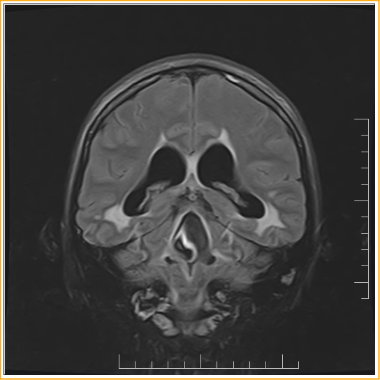
Obstruction of Magendie's Foramen :MRI
Case Report :
A 33-year-old woman presented with visual disturbance and balance difficulty on MRI brain shows dilatation of lateral, III and IV ventricles along with periventricular T2/FLAIR hyperintensity. Temporal horns are dilated. Fourth ventricle appears dilated out of propotion along with prominent CSF flow void in the IV ventricle and some enhancement in ependymal surface of IV ventricle. Contour abnormality in the foramen of magendie. These findings are indicative of IV ventricular outflow tract obstruction possibly post infective sequele/arachnoiditis in foramen of magendie
Teaching Points:
- Membranous obstruction of the Magendie's foramen is a rare case of non communicating quadriventricular hydrocephalus. In children is usually congenital and related with Dandy-Walker Syndrome, Arnold-Chiari malformation, tuberous sclerosis, spina bifida, platybasia, achondroplasia, basilar impression and atlanto-occipital fusion.
- In adults it is mostly acquired rather than congenital. Acquired ventricuΙar outlet obstructions are reported in adults as well as children and generally occur in infection (meningoencephalitides, prenatal infection, shunting procedures, granulomatosis, venereal disease, influenza, ear-ocular-nasopharyngeal infection, Toxoplasmosis, Cysticercosis), head trauma, intraventricular hemorrhage, tumors or Arnold-Chiari malformation
Obstruction of Magendie's Foramen :MRI
 Reviewed by Sumer Sethi
on
Thursday, September 07, 2017
Rating:
 Reviewed by Sumer Sethi
on
Thursday, September 07, 2017
Rating:


No comments:
Post a Comment