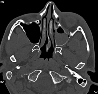
Blow Out Fracture-MDCT
A young adult with history of injury to the left face with diplopia and restricted gaze, shows on the multidetector CT (128 slices), an orbital floor fracture with inferior rectus intrapment with a chip of the communited component lying as a satellite piece.
Teaching points by Dr MGK Murthy, Dr Sumer Sethi, Mr Hari Om & Mr Shekhar:
Smith and Regan described blowout fracture in 1957, as orbital floor fracture (maxillary / zygomatic / palatine bones constitute the floor measuring 35 to 40 mm and shortest of all the walls, not reaching upto the orbital apex). Second most common mid facial fracture after nasal bones.
Mechanism usually involves impact injury, large enough not to perforate the globe and small enough not to fracture orbital rim.
Leads to increase in intraorbital hydrolic pressure.
Most occur in posteromedial region which is the thinnest and medial to the infraorbital groove.
Orbital emphysema, intraorbital bleed, inferior rectus entrapment, globe injuries including Hyphaema, retinal injury are possible accompaniments.
CT delineates the fracture including any associated fractures like zygomatic / lefort type II / III, and fracture orbital rims.
Early repair with plating and steel wiring has been practiced for long along with recent biocompatible implants and microplatings.
Blow Out Fracture-MDCT
 Reviewed by Sumer Sethi
on
Friday, November 18, 2011
Rating:
 Reviewed by Sumer Sethi
on
Friday, November 18, 2011
Rating:


No comments:
Post a Comment